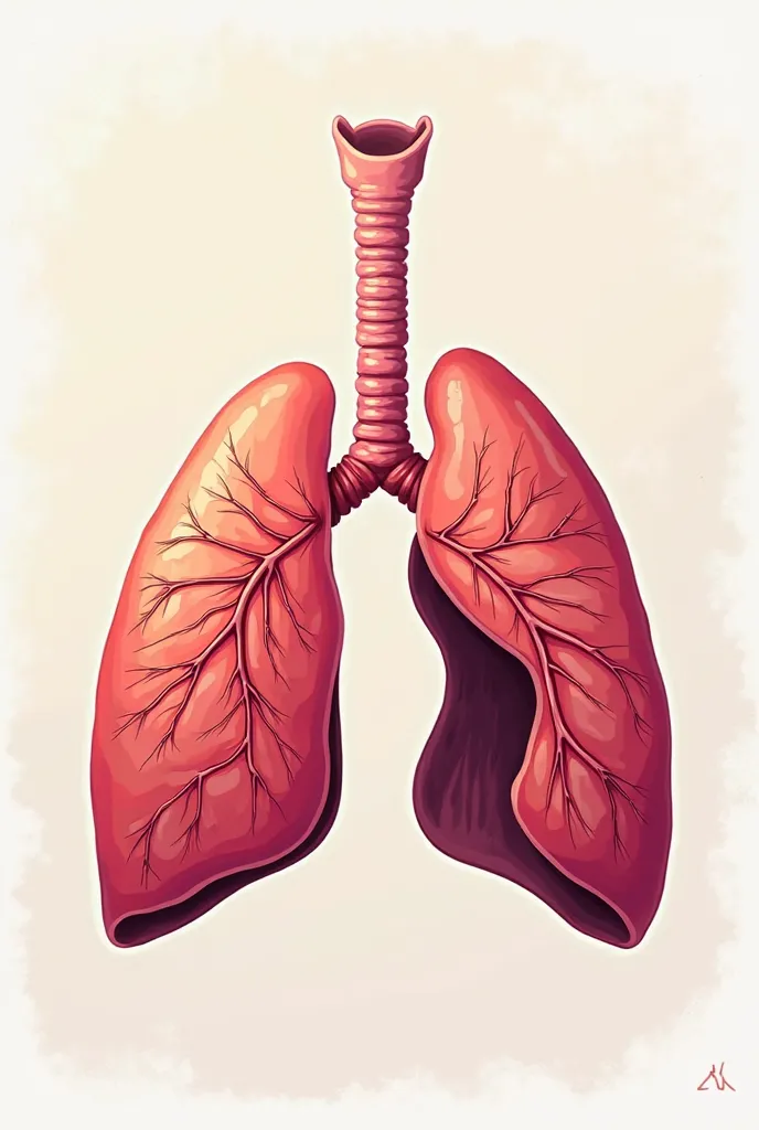
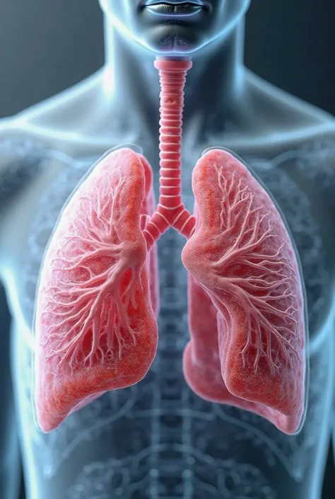
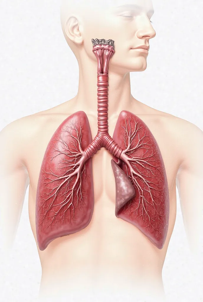
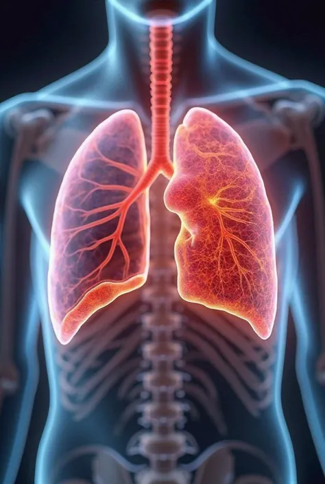
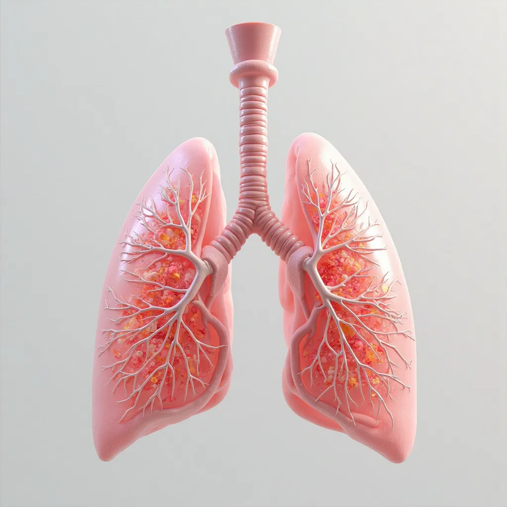
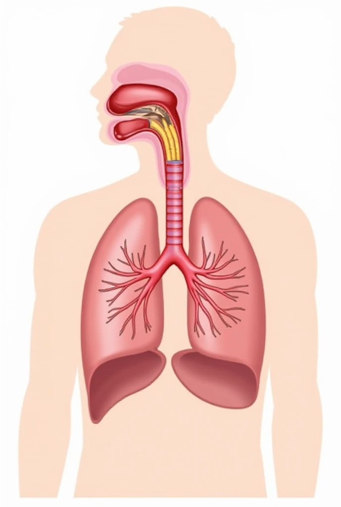
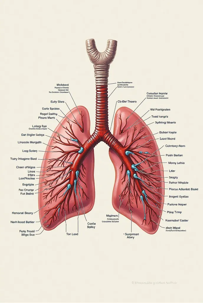
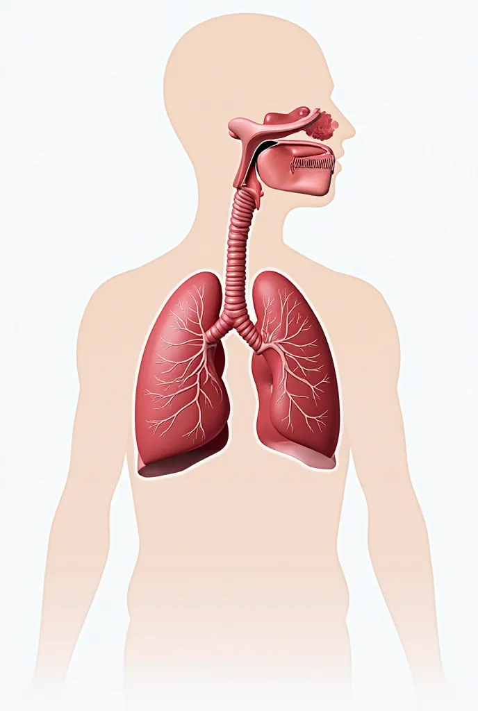
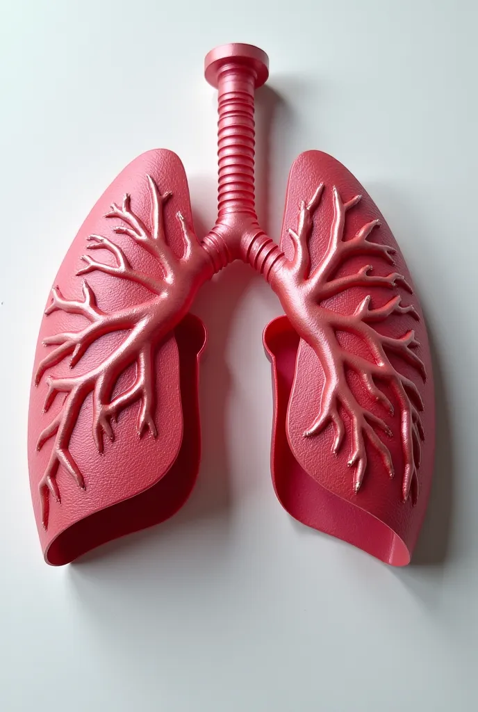
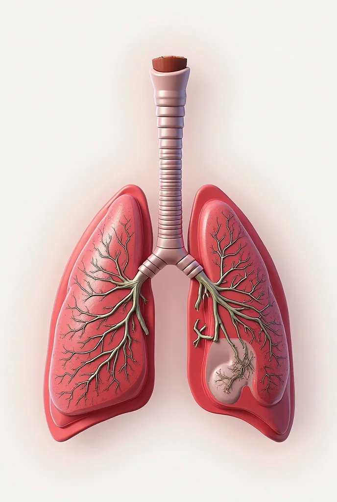
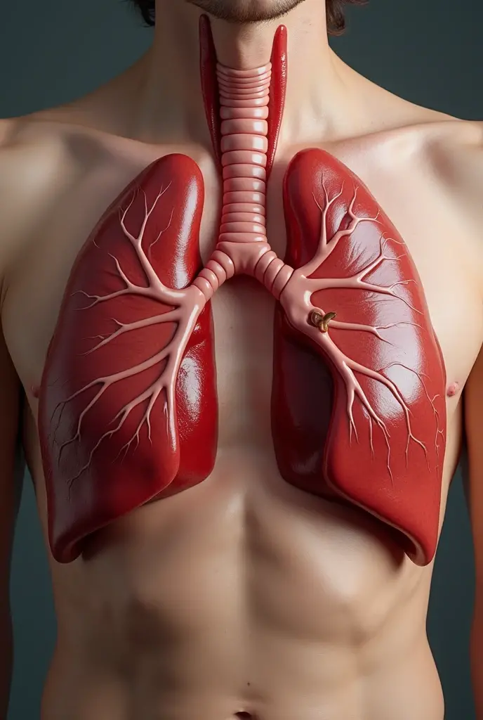
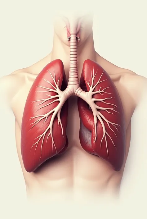
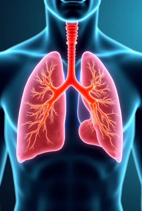
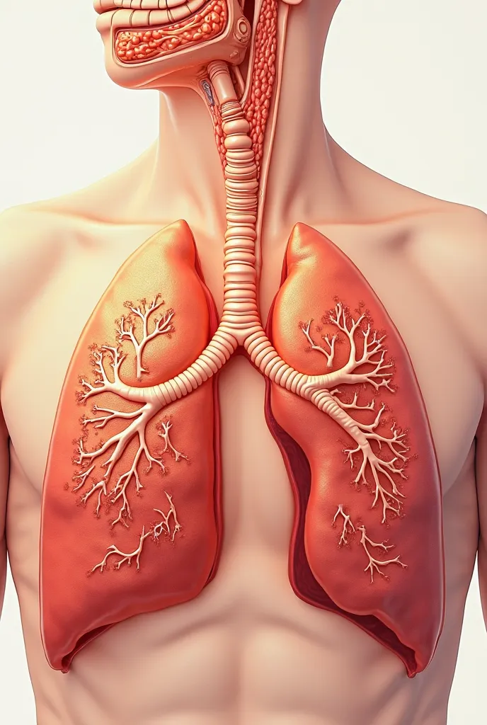
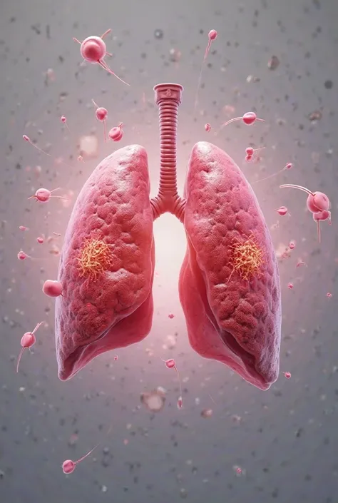
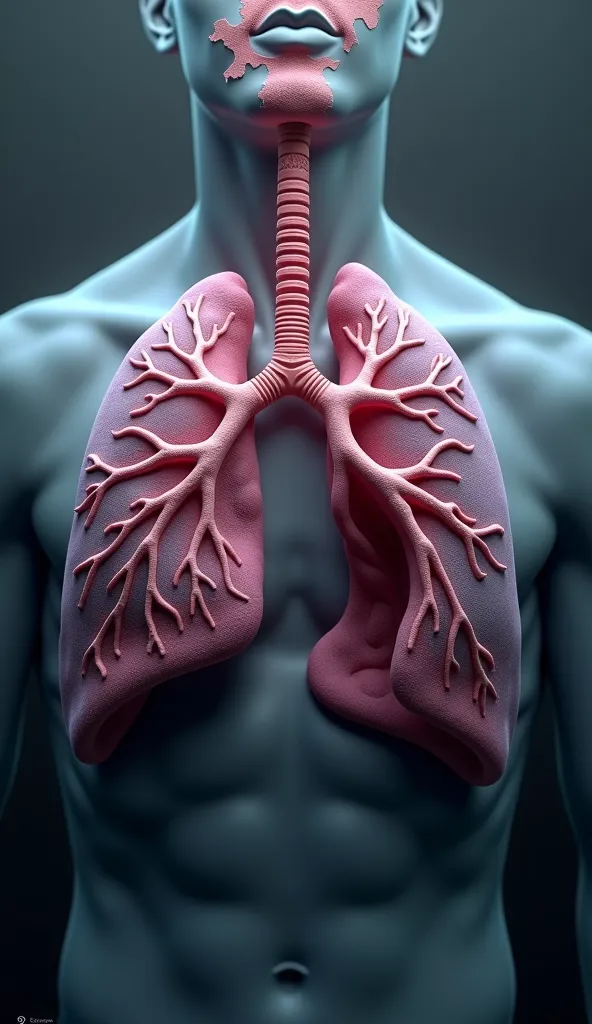
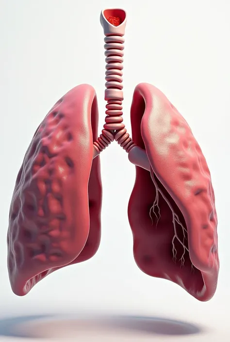
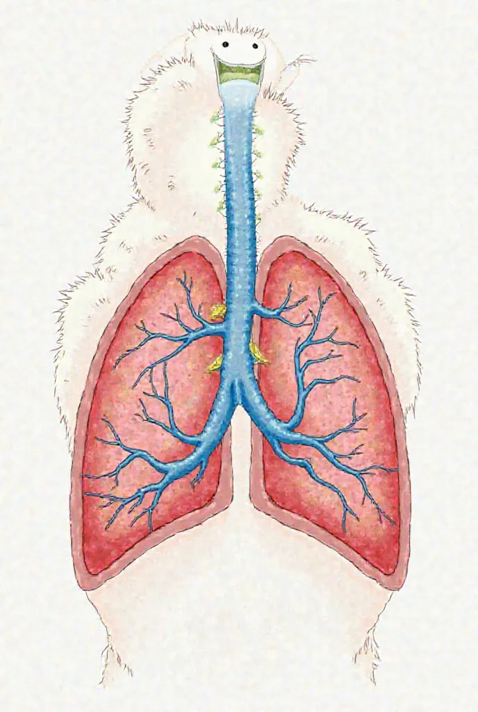
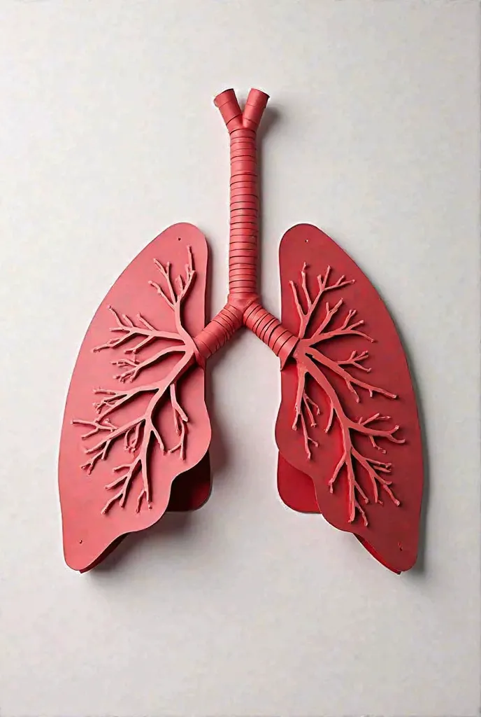
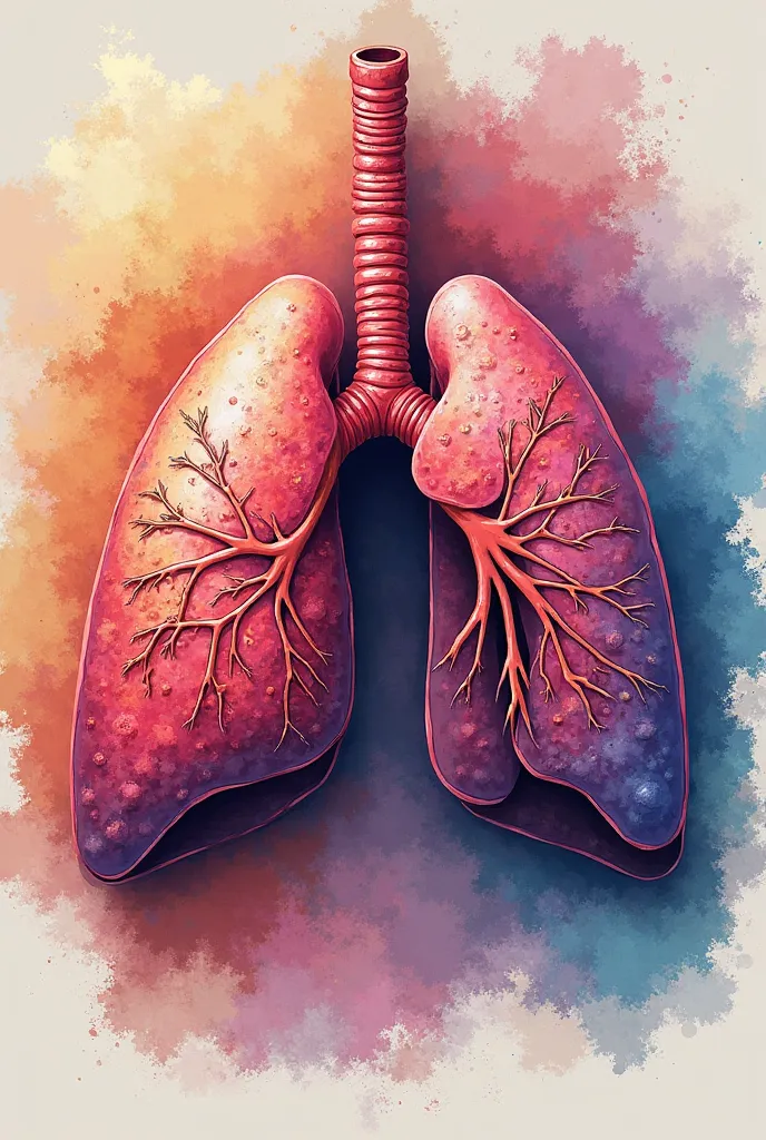
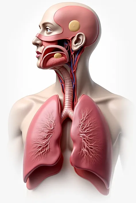
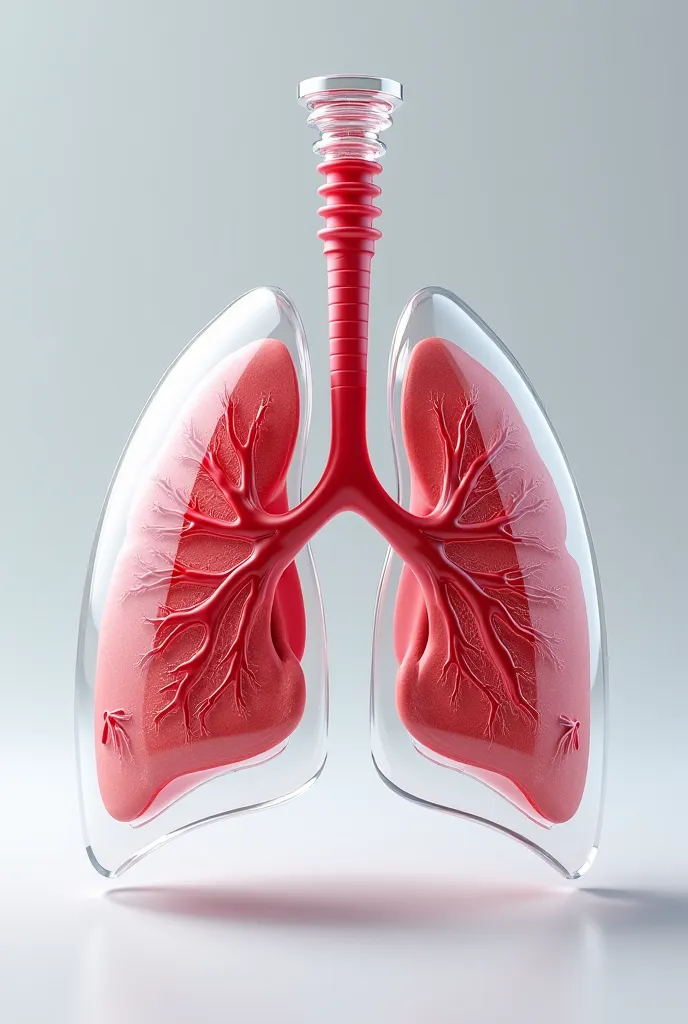
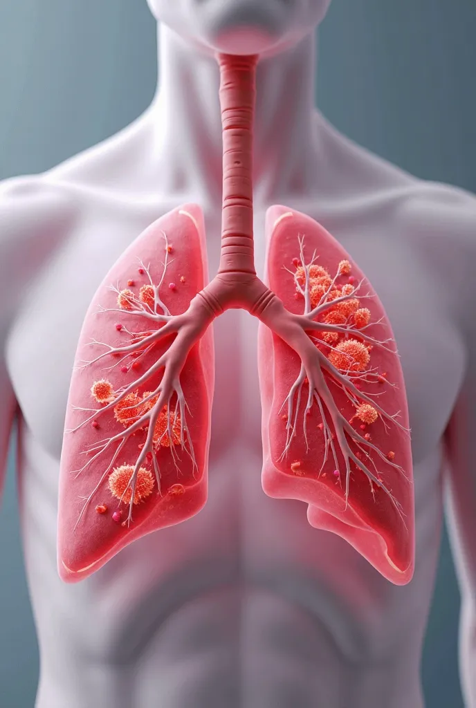
Create a highly detailed and educational illustration of a human lung
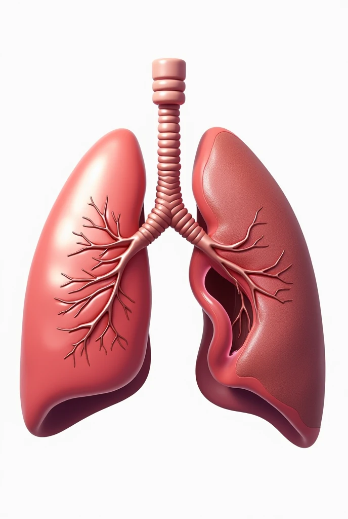


Create a highly detailed and educational illustration of a human lung, oriented to fit a poster with dimensions 30x24 inches. The image should be split to show a clear comparison between two lobes of the lung:Normal Lobe: On one side, depict a healthy lung lobe with its anatomy clearly visible. Illustrate the bronchi, alveoli, and blood vessels with normal, unobstructed airways and tissue. Use realistic colors and textures to represent the healthy tissue's pinkish hue and smooth, clean appearance.COPD-Affected Lobe: On the other side, show a lung lobe affected by Chronic Obstructive Pulmonary Disease (COPD). Highlight the key pathological changes, including:Emphysema: Illustrate enlarged alveoli and a loss of alveolar walls, showing the resulting larger, air-filled spaces and reduced surface area for gas exchange.Chronic Bronchitis: Depict thickened bronchial walls, increased mucus production, and potential inflammation of the airways.Air Trapping: Show areas of the lung with trapped air and less effective air movement.Include labels and annotations to identify and explain the normal and affected features. Use contrasting colors and textures to clearly differentiate between the healthy and pathological areas. The overall style should be realistic and informative, suitable for educational purposes and medical presentations
Generation Data
Records
Prompts
Copy
Create a highly detailed and educational illustration of a human lung
,
oriented to fit a poster with dimensions 30x24 inches
.
The image should be split to show a clear comparison between two lobes of the lung:Normal Lobe: On one side
,
depict a healthy lung lobe with its anatomy clearly visible
.
Illustrate the bronchi
,
alveoli
,
and blood vessels with normal
,
unobstructed airways and tissue
.
Use realistic colors and textures to represent the healthy tissue's pinkish hue and smooth
,
clean appearance
.
COPD-Affected Lobe: On the other side
,
show a lung lobe affected by Chronic Obstructive Pulmonary Disease (COPD)
.
Highlight the key pathological changes
,
including:Emphysema: Illustrate enlarged alveoli and a loss of alveolar walls
,
showing the resulting larger
,
air-filled spaces and reduced surface area for gas exchange
.
Chronic Bronchitis: Depict thickened bronchial walls
,
increased mucus production
,
and potential inflammation of the airways
.
Air Trapping: Show areas of the lung with trapped air and less effective air movement
.
Include labels and annotations to identify and explain the normal and affected features
.
Use contrasting colors and textures to clearly differentiate between the healthy and pathological areas
.
The overall style should be realistic and informative
,
suitable for educational purposes and medical presentations
INFO
Checkpoint & LoRA

Checkpoint
SeaArt Infinity
#SeaArt Infinity
0 comment
1
0
0
SeaArt Swift AI Apps

AI Video Generation
Unleash your imagination and let AI create visual wonders for you

Face Swap Online Free
Create funny or realistic face swap videos & photos in a snap

Anime to Reality
Instantly bring your favorite anime characters to life.

AI Eraser
Easily remove unwanted objects, watermarks, or people from your photos.

AI Dance Video Generator
Play with this AI dance video generator, unleash your inner dancer instantly!

AI Filters
Turns every photo into a work of art
Explore More AI Apps